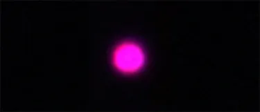
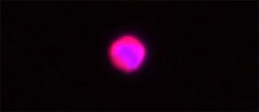
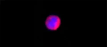
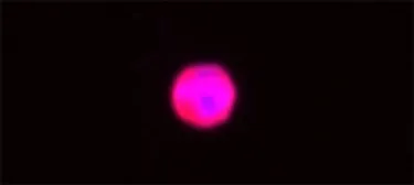
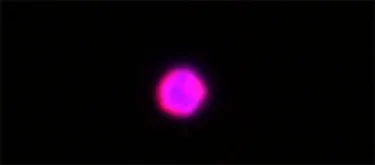
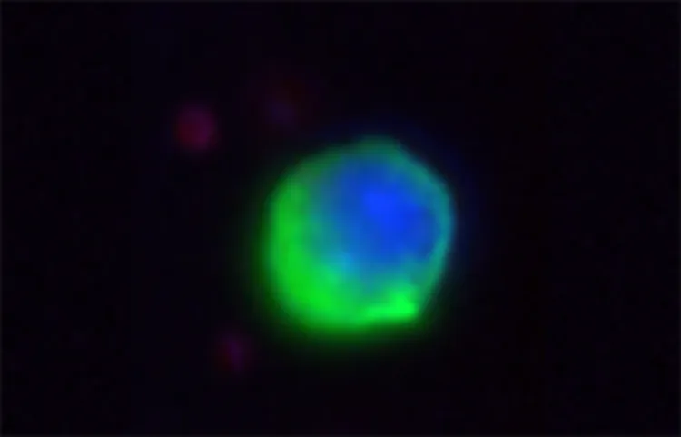
CTC Atlas
The CTC Atlas is the first open-access and potentially groundbreaking resource that combines advanced molecular imaging techniques and meticulous sample preparation to offer unparalleled insights into circulating tumor cells (CTCs) and other tumor-related cells. Developed thanks to the funding of Ministry of Science and Higher Education of Poland, and based on the newest technologies such as imaging flow cytometry, the Atlas provides high-quality visual and analytical data, empowering researchers and clinicians to better understand cancer biology. Simply scroll through the gallery of images below and click on any image to view a detailed description of its features and methodology.
Methodology Behind the Atlas
The biological material for analysis is derived from tumor draining or peripheral blood of oncological patients recruited after informed written consent according to protocols approved by the Independent Bioethics Committee for Scientific Research at Medical University of Gdańsk. Blood is collected to EDTA tubes, and processed as soon as possible after blood collection (optimally within 3 hours after donation). First blood draw is discarded to avoid skin cell (i.e., keratinocytes, fibroblasts) and endothelial cell contamination during the puncture. Cells are isolated using density gradient centrifugation, yielding a mononuclear blood cell fraction containing, among others, putative circulating tumor cells (CTCs). These cells are then fixed in 4% formaldehyde, and stored at −80 °C for further analysis.
Prepared samples are stained using different multimarker immunofluorescence protocols containing both CTC inclusion markers (e.g. pan-keratin) and at least 2 exclusion markers (at least CD45 and CD31). The investigated cells are thawed and washed with 1xPBS to remove the formaldehyde, and then incubated with an antibody cocktail. After one additional washing step in 1xPBS, the cells were resuspended in 1xPBS, counterstained with DAPI, and immediately analyzed using the imaging flow cytometer (i.e. Amnis® ImageStream® X Mk II, Cytec Biosciences).
The Atlas is continuously being expanded and updated with new images and data containing new features of identified cells and new protocols including further markers, making it a valuable resource for scientists and physicians. Users can browse the Atlas to explore images categorized by cell types and their features, and gain insights into their identification methods and potential diagnostic significance.
More detailed information on specific staining protocols for CTCs and other tumor-derived cells from various cancers can be found in the following publications. We kindly ask CTC Atlas users to cite the following publications when referening to our data:
For pan-K/vim/DAPI/CD45/CD31/α-SMA/CD29 protocol and CTC phenotyping in breast cancer:
Muchlińska Anna, Wenta Robert, Ścińska Wiktoria, Markiewicz Aleksandra, Suchodolska Grażyna, Senkus Elżbieta, Żaczek Anna, Bednarz-Knoll Natalia. Improved characterization of circulating tumor cells and cancer-associated fibroblasts in one-tube assay in breast cancer patients using imaging flow cytometry. Cancers 2023, 15(16): 4169 (doi: 10.3390/cancers15164169)
For imaging flow cytometry implementation in liquid biopsy:
Muchlińska Anna, Smentoch Julia, Żaczek Anna J, Bednarz-Knoll Natalia. Detection and Characterization of Circulating Tumor Cells Using Imaging Flow Cytometry—A Perspective Study. Cancers 2022, 14(17): 4178 (doi: 10.3390/cancers14174178)
For measurable morphological features of CTCs:
Wenta Robert, Richert Julia, Muchlińska Anna, Senkus Elżbieta, Suchodolska Grażyna, Łapińska-Szumczyk Sylwia, Domżalski Paweł, Miszewski Kevin, Matuszewski Marcin, Dziadziuszko Rafał, Supernat Anna, Żaczek Anna, Bednarz-Knoll Natalia. Measurable morphological features of single circulating tumor cells in selected solid tumors—A pilot study. Cytometry 2024, 105(12):883-892 (doi: 10.1002/cyto.a.24906)